1. Compare the structure of the atria and ventricles - How are they different? Why is that?
Ventricles are thick walled muscular, which is essential because they must be strong enough to to push blood away from the heart and through the body. The atria are thinner because blood flows into the atria, and very little force is needed to move blood through the atria.
2. Did you notice a difference between the veins and the arteries entering and leaving the heart? How is their structure different?
The pulmonary arteries carry blood from the heart to the lungs. Pulmonary veins carry blood from the lungs to the heart. Arteries have thick walls, and veins have thinner walls. Other than that, we did not notice much of a difference.
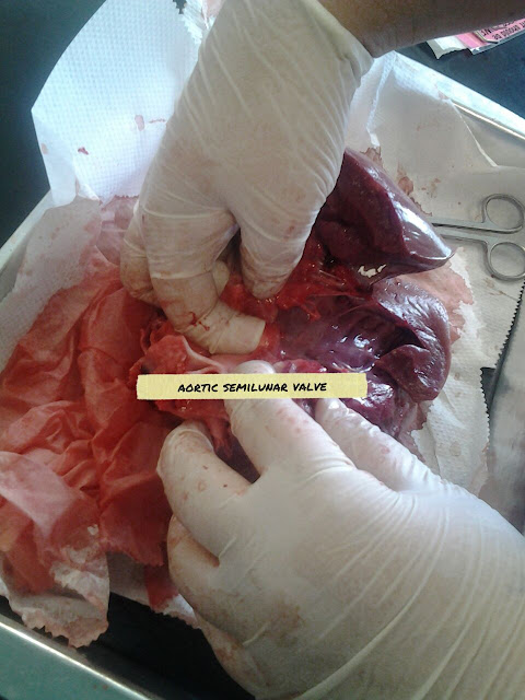
3. Describe the valves that you found in the heart - what are their functions?
The valves we found are the aortic semilunar valves, and the pulmonary semilunar valves. The blood flows through the semilunar valves on its way out of the heart. The right ventricle has a pulmonary semilunar valve, since it pumps blood out through the pulmonary artery. The left side has an aortic semilunar valve, since it pumps out blood through the aorta. They prevent blood from flowing back into the ventricles.
4. What surprised you about dissecting the heart? Why?
What surprised me is how similar it was to a human heart. The pictures and diagrams we have seen in class match the heart we dissected. I was expecting some differences.


No comments:
Post a Comment