I may not seem very engaged in class, but I promise, I am...at least most of the time >-< It is sometimes difficult to say things because the louder kids often beat me to it, but that's all right. I have discovered that the best way to be engaged in class is by maintaining eye contact with the teacher. Eye contact makes it easier to focus and not get distracted. I really enjoyed the question box, because it gave us all a chance to learn about something we have been curious about. Engagement also means to be in class on time, with all the necessary supplies. I would say I have done a good job of staying engaged in Biology 12.
Why is the grass green?
Monday, 3 June 2013
Tuesday, 14 May 2013
Space and URINE!
I'll bet you a nickel that you've never stopped to think about how astronauts accommodate the needs of their urinary system while on a mission. What a weird thing to wonder, right? Well, the following post will enlighten your noggin.
Both male and female astronauts urinate into a funnel, nicknamed Mr. Thirsty, which is attached to a tube. A gentle vacuum then sucks the urine into a tube without spilling a drop. When the tank is full, it shoots the urine outside, where it freezes into clouds of ice crystals that look like stars. Astronaut Wally Schirra liked to call it "Constellation Urion."
The International Space Station is building a system to purify and reuse the water in urine, of both humans and animals. NASA estimates that 72 rats urinate about as much as one astronaut.
Source #1 Source #2
Also, here's a random bonus urine related fact:
Thursday, 9 May 2013
Circulatory System Quiz Review
The Pulmonary system carries deoxygenated blood out from the pulmonary arteries to the lungs, so that it can become oxygenated. Oxygenated blood then comes back from the lungs through the pulmonary veins, and into the left atrium. From there, the systemic system comes into play. The systemic system carries oxygenated blood to the rest of the body. Arteries are thick-walled, and carry oxygenated blood throughout the body. Veins are thinner-walled, and carry deoxygenated blood throughout the body; they are thicker in diameter and contain many valves, to prevent blood from flowing backward due to gravitational forces. Pulmonary arteries carry deoxygenated blood blood; pulmonary veins carry oxygenated blood.
Blood flow from the common carotid artery to the aorta:
common carotid artery -> brain -> jugular vein -> superior vena cava -> right atrium -> av (tricuspid) valve -> right ventricle -> pulmonary semilunar valve -> pulmonary trunk -> pulmonary arteries -> lungs -> pulmonary veins -> left atrium -> av (bicuspid/mitral) valve -> left ventricle -> aortic semilunar valve -> aorta
Three major modifications of the fetal circulation system are the foramenovale, ductus arteriosis, and ductus venosus. The foramenovale is a hole in the heart, located in between the atria; blood flows through this hole to bypass the lungs, as a fetus is not required to breathe. The ductus arteriosis is a hole in between the pulmonary trunk and aorta, which is also for blood to bypass the lungs. The ductus venosus is a channel in the fetus, joining the umbilical vein to the inferior vena cava; it carries oxygen rich blood from the placenta to the baybee.
Three major modifications of the fetal circulation system are the foramenovale, ductus arteriosis, and ductus venosus. The foramenovale is a hole in the heart, located in between the atria; blood flows through this hole to bypass the lungs, as a fetus is not required to breathe. The ductus arteriosis is a hole in between the pulmonary trunk and aorta, which is also for blood to bypass the lungs. The ductus venosus is a channel in the fetus, joining the umbilical vein to the inferior vena cava; it carries oxygen rich blood from the placenta to the baybee.
Wednesday, 1 May 2013
Playland!
So, today turned out to be way more fun than I had expected. We went on some rides, ate some food, and soaked in some sun. Good times, good times. I couldn't take many good pictures because it was bright, and everything was moving, but here are the ones that turned out quite well.
Thanks for organizing this trip, Ms. Phillips!
Thursday, 25 April 2013
Heart Dissection
1. Compare the structure of the atria and ventricles - How are they different? Why is that?
Ventricles are thick walled muscular, which is essential because they must be strong enough to to push blood away from the heart and through the body. The atria are thinner because blood flows into the atria, and very little force is needed to move blood through the atria.
2. Did you notice a difference between the veins and the arteries entering and leaving the heart? How is their structure different?
The pulmonary arteries carry blood from the heart to the lungs. Pulmonary veins carry blood from the lungs to the heart. Arteries have thick walls, and veins have thinner walls. Other than that, we did not notice much of a difference.
3. Describe the valves that you found in the heart - what are their functions?
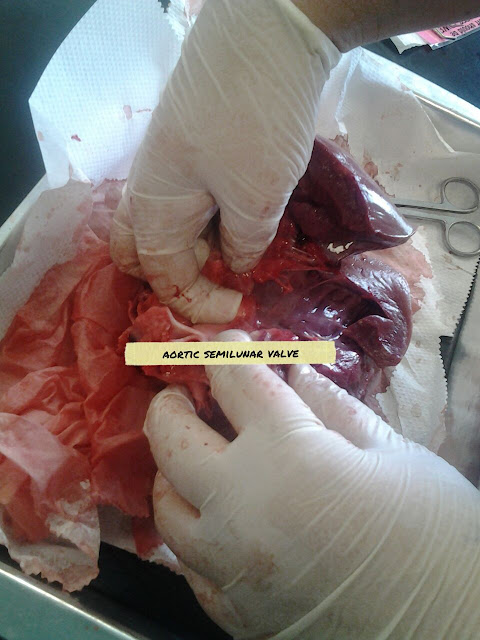
The valves we found are the aortic semilunar valves, and the pulmonary semilunar valves. The blood flows through the semilunar valves on its way out of the heart. The right ventricle has a pulmonary semilunar valve, since it pumps blood out through the pulmonary artery. The left side has an aortic semilunar valve, since it pumps out blood through the aorta. They prevent blood from flowing back into the ventricles.
4. What surprised you about dissecting the heart? Why?
What surprised me is how similar it was to a human heart. The pictures and diagrams we have seen in class match the heart we dissected. I was expecting some differences.
How much blood is pumped by the heart?
As we all know, the heart is the strongest muscle in our body, and is also the most vital. The million dollar question is: how much blood does the heart pump?
The average human heart beats :
- 72 times a minute
- 100, 000 times a day
- 3, 600, 000 times a year
- 2.5 billion times a lifetime
- A healthy human heart pumps 2, 000 gallons of blood through 60, 000 miles of blood vessels every day.
- A kitchen faucet would need to be turned on all the way for at least 45 years to equal the amount of blood pumped in an average lifetime.
- The volume of blood pumped by the heart can range from five to 30 litres per minute.
- The heart creates enough energy, per day, to drive a truck 20 miles. That is equal to driving to the the moon and back in a lifetime.
- During an average lifetime, the heart will pump about 1.5 barrels of blood, which is enough to fill 200 train tank cars.
Wednesday, 24 April 2013
Subscribe to:
Comments (Atom)


