I may not seem very engaged in class, but I promise, I am...at least most of the time >-< It is sometimes difficult to say things because the louder kids often beat me to it, but that's all right. I have discovered that the best way to be engaged in class is by maintaining eye contact with the teacher. Eye contact makes it easier to focus and not get distracted. I really enjoyed the question box, because it gave us all a chance to learn about something we have been curious about. Engagement also means to be in class on time, with all the necessary supplies. I would say I have done a good job of staying engaged in Biology 12.
Monday, 3 June 2013
Tuesday, 14 May 2013
Space and URINE!
I'll bet you a nickel that you've never stopped to think about how astronauts accommodate the needs of their urinary system while on a mission. What a weird thing to wonder, right? Well, the following post will enlighten your noggin.
Both male and female astronauts urinate into a funnel, nicknamed Mr. Thirsty, which is attached to a tube. A gentle vacuum then sucks the urine into a tube without spilling a drop. When the tank is full, it shoots the urine outside, where it freezes into clouds of ice crystals that look like stars. Astronaut Wally Schirra liked to call it "Constellation Urion."
The International Space Station is building a system to purify and reuse the water in urine, of both humans and animals. NASA estimates that 72 rats urinate about as much as one astronaut.
Source #1 Source #2
Also, here's a random bonus urine related fact:


Thursday, 9 May 2013
Circulatory System Quiz Review


The Pulmonary system carries deoxygenated blood out from the pulmonary arteries to the lungs, so that it can become oxygenated. Oxygenated blood then comes back from the lungs through the pulmonary veins, and into the left atrium. From there, the systemic system comes into play. The systemic system carries oxygenated blood to the rest of the body. Arteries are thick-walled, and carry oxygenated blood throughout the body. Veins are thinner-walled, and carry deoxygenated blood throughout the body; they are thicker in diameter and contain many valves, to prevent blood from flowing backward due to gravitational forces. Pulmonary arteries carry deoxygenated blood blood; pulmonary veins carry oxygenated blood.
Blood flow from the common carotid artery to the aorta:
common carotid artery -> brain -> jugular vein -> superior vena cava -> right atrium -> av (tricuspid) valve -> right ventricle -> pulmonary semilunar valve -> pulmonary trunk -> pulmonary arteries -> lungs -> pulmonary veins -> left atrium -> av (bicuspid/mitral) valve -> left ventricle -> aortic semilunar valve -> aorta
Three major modifications of the fetal circulation system are the foramenovale, ductus arteriosis, and ductus venosus. The foramenovale is a hole in the heart, located in between the atria; blood flows through this hole to bypass the lungs, as a fetus is not required to breathe. The ductus arteriosis is a hole in between the pulmonary trunk and aorta, which is also for blood to bypass the lungs. The ductus venosus is a channel in the fetus, joining the umbilical vein to the inferior vena cava; it carries oxygen rich blood from the placenta to the baybee.


Three major modifications of the fetal circulation system are the foramenovale, ductus arteriosis, and ductus venosus. The foramenovale is a hole in the heart, located in between the atria; blood flows through this hole to bypass the lungs, as a fetus is not required to breathe. The ductus arteriosis is a hole in between the pulmonary trunk and aorta, which is also for blood to bypass the lungs. The ductus venosus is a channel in the fetus, joining the umbilical vein to the inferior vena cava; it carries oxygen rich blood from the placenta to the baybee.

Wednesday, 1 May 2013
Playland!
So, today turned out to be way more fun than I had expected. We went on some rides, ate some food, and soaked in some sun. Good times, good times. I couldn't take many good pictures because it was bright, and everything was moving, but here are the ones that turned out quite well.
Thanks for organizing this trip, Ms. Phillips!


Thursday, 25 April 2013
Heart Dissection


1. Compare the structure of the atria and ventricles - How are they different? Why is that?
Ventricles are thick walled muscular, which is essential because they must be strong enough to to push blood away from the heart and through the body. The atria are thinner because blood flows into the atria, and very little force is needed to move blood through the atria.
2. Did you notice a difference between the veins and the arteries entering and leaving the heart? How is their structure different?
The pulmonary arteries carry blood from the heart to the lungs. Pulmonary veins carry blood from the lungs to the heart. Arteries have thick walls, and veins have thinner walls. Other than that, we did not notice much of a difference.
3. Describe the valves that you found in the heart - what are their functions?
The valves we found are the aortic semilunar valves, and the pulmonary semilunar valves. The blood flows through the semilunar valves on its way out of the heart. The right ventricle has a pulmonary semilunar valve, since it pumps blood out through the pulmonary artery. The left side has an aortic semilunar valve, since it pumps out blood through the aorta. They prevent blood from flowing back into the ventricles.
4. What surprised you about dissecting the heart? Why?
What surprised me is how similar it was to a human heart. The pictures and diagrams we have seen in class match the heart we dissected. I was expecting some differences.


How much blood is pumped by the heart?
As we all know, the heart is the strongest muscle in our body, and is also the most vital. The million dollar question is: how much blood does the heart pump?
The average human heart beats :
- 72 times a minute
- 100, 000 times a day
- 3, 600, 000 times a year
- 2.5 billion times a lifetime
- A healthy human heart pumps 2, 000 gallons of blood through 60, 000 miles of blood vessels every day.
- A kitchen faucet would need to be turned on all the way for at least 45 years to equal the amount of blood pumped in an average lifetime.
- The volume of blood pumped by the heart can range from five to 30 litres per minute.
- The heart creates enough energy, per day, to drive a truck 20 miles. That is equal to driving to the the moon and back in a lifetime.
- During an average lifetime, the heart will pump about 1.5 barrels of blood, which is enough to fill 200 train tank cars.


Wednesday, 24 April 2013


Friday, 5 April 2013
What is Asthma?
Asthma is a respiratory condition marked by spasms of the bronchi in the lungs, which causes difficulties in breathing. Asthma causes recurring periods of wheezing, chest tightness, shortness of breath, and coughing. The coughing often occurs at night or early in the morning. People who have asthma have inflamed airways, making them swollen and very sensitive; they tend to react strongly to certain inhaled substances. When the airways react, the muscles around them tighten. This narrows the airways, causing less air to flow into the lungs. Cells in the airways might produce more mucus than usual; this chain reaction can result in asthma symptoms. Symptoms can happen each time the airways are inflamed.
There are many factors which contribute to asthma, such as:
- low birth weight
- exposure to tobacco smoke
- allergies
- pollution
- pregnancy
- stress
- genes
- airway hyperactivity
- atopy
Some triggers:
- mould
- animal dander
- pollen
- cockroaches
- dust mites
- chemicals, fumes, odours
- respiratory viral infections
- weather
- smog
- excessive, strenuous physical activity
Although asthma cannot be cured, it can be controlled by avoiding asthma triggers, taking medication, and following an asthma action plan.


Vital Capacity
We tested our vital capacity earlier this week and I can store up to 2800 cc or 2.8 L. I am happy with this result, as 2.7 L is the average for a 17 year old girl. Vital capacity is what it is depending on how much we use our lungs. Some of the highest vital capacities out of the girls were from those in band; the band kids had higher lung capacities because they have conditioned and strengthened their lungs. A usual breath consists of 500 mL. The reason we do not exhale our entire vital capacity each time is to conserve energy; it would be exhausting to force out all the air each time we need to exhale. The purpose of exhaling is to rid the body of carbon dioxide to prevent our blood from becoming toxic. Breathing is meant to be an autonomic process to allow us to concentrate on other things. Activities such as playing a sport or an instrument are beneficial to our lungs because it allows them to become stronger. The lungs are like any other muscle in our body, they become stronger the more they are exercised.


Friday, 15 March 2013
Hey, look how much fun respiration is!
Today we made lungs one singular lung.
*I wonder if we could implant it into a real person.*
*coughcough*
Nope, never mind.
Bad idea.


*I wonder if we could implant it into a real person.*
*coughcough*
Nope, never mind.
Bad idea.
Look at this super artistic picture I took. YEAH.
Our first attempt wasn't working, so we tried again.
Here's what it looks like in action:

So, we learned that the contraction of the diaphragm (bottom balloon) is what happens when we inhale, and it relaxes when we exhale. When we breathe, the ribcage (the plastic cup) expands to allow the lungs (balloon inside the cup) enough room to inflate. When the diaphragm contracts, air rushes through the trachea (straw) into the lungs. They deflate when the diaphragm goes back into it's resting position. Basically, the *breathing in* part requires a muscle contraction of the diaphragm.


Thursday, 14 March 2013
Unit 1 Review
Hey there, Bio ..

Water


Water
- breaks down nutrients
- is stuck together by cohesive, polar covalent hydrogen bonds
- helps the body get rid of waste (as a components of urine and sweat)
- regulates our temperature because it has a high specific heat capacity
- lubricates joints
- is a universal solvent
- protects our brains
- is polar, which allows hydrogen bonding
Digestive System
Oh boy, I sure hope these show up.
Sorry for being lazy D:
This, basically, is the terms.
- Swallowing is the act of pushing the bolus of food down into the esophagus. Peristalsis is what acts upon the food from this point on; it is the name of the contractions of the muscle layers present in the digestive tract.
- The pancreas is the source gland for insulin. Insulin maintains blood glucose levels by either up taking the intake of glucose, storing it, or releasing it into the blood.
- The liver produces bile, stores glucose as glycogen, detoxifies alcohol and toxins from blood, processes nutrients from small intestine, produces blood proteins, and converts amonia in to urea.
- Bile emulsifies fats by breaking them down so they can be digested and absorbed as easily as possible.
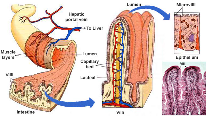
- The small intestine is equipped for both chemical and physical digestion. Physical digestion is done by peristalsis, and the villi and microvilli. The small intestine receives enzymes and secretions from the pancreas to aid in chemical digestion.
- Villi and microvilli are present in the small intestine; their purpose is to maximize nutrient absorption. Glucose and amino acids are absorbed into the capillaries within the villi, are taken into the blood stream, and are sent to the liver. Fatty acids and glycerol are absorbed into the lacteal of the villi, and travel in the lymphatic system.
- The anaerobic bacteria in the colon prevents the growth of harmful bacteria. By taking up space in the colon, it limits the growth of pathogenic bacteria .
- Water helps break down molecules into their monomers.
- Sodium bicarbonate is released into the small intestine by the pancreas so the gastric juice does not burn the duodenum lining.
- HCl in the stomach is essential in killing harmful bacteria, so that it is not absorbed into the body. It also changes the pH of salivary amylase, rendering it useless.
- Mucus protects stomach lining from the wrath of HCl.
Biological Molecules
There's a review booklet.
Oh hey, maybe I actually get this stuff..


Wednesday, 6 March 2013
Biology 12 Interim Report
Every day in this class is a chance to feel accomplished; I especially felt successful when I realized that I am able to answer questions without having to go through my notes. It is comforting to know that I do not have to flail for answers, they are in my brain.
My work habits are great. When I go through my binder, there are no incomplete worksheets. I have submitted all my assignments and hope to continue to do so in the future.
I have had a chance to work collaboratively with some of my classmates. It has enhanced my learning in the sense that I learn more effectively with other people. If one of my group members has a question that had not come to my mind, I get a chance to learn at least one more thing than I would have on my own.
My main goal is to get an A in order to graduate with a 4.0 average. To do so, I need to study more effectively. By getting a high mark I will have proven to myself that I have learned and applied new concepts. Learning is fun, but is often overshadowed by the anxiety of getting a high mark on tests and projects. I hope to be able to balance all the aspects in order to gain a holistic experience. More than anything, I want to have fun in Biology 12.
Biology is A W E S O M E.

Monday, 25 February 2013
Biochemical Molecules Test

1. We used a variety of foods for this test, namely tomato, cheese, cupcake, lettuce, bread, apple, orange, carrot, and pancake. For the simple sugars test, tomato and carrot tested positive.


Cupcake, bread, and pancake tested positive for starch; tomato and cheese had a few specs of black which showed the reaction between the starch and iodine.

Butter, cupcake, and bread tested positive for lipids.

2. The building blocks of starch molecules is glucose. Glucose polymers are made up of Amylose and Amylopectin. When water is added, starch breaks down into glucose molecules. The following is an image of the molecular structure of starch (I hope D:):

3. Simple carbohydrates digest or break down into sugar quickly. The reason some test tubes needed to remain in the hot water bath longer before turning a cloudy orange or yellow, is because the more complex a carbohydrate is, the longer it takes to break down.
4. Starting at the mouth, using our teeth and tongue, we chew up food to increase surface area, therefore making the process of digestion slightly easier. As we chew, salivary amylase is secreted, which digests complex carbohydrates. The bolus of food is then sent to the back of the mouth, through the pharynx, and into the esophagus. The bolus makes its way down to the stomach through muscle contractions called peristalsis, which can be found throughout the entire digestive tract. Upon making it through the cardiac sphincter, and reaching the stomach, the bolus experiences more mechanical digestion, along with chemical digestion, with the help of gastric juice, composed of hydrochloric acid, mucus and the enzyme pepsin. The pH inside the stomach is 2, which is why mucus is an essential component of the stomach; it lines the stomach walls to prevent corrosion due to the HCl. Proteins begin to digest in the stomach. The food (chyme) then passes through the pyloric sphincter, into the duodenum. This is where the accessory organs, the liver, gall bladder, and pancreas, come into play. The pancreas has pancreatic juice, containing pancreatic amylase, trypsin, lipase, nuclease, and sodium bicarbonate; it also produces insulin and glucagon. The liver produces bile, and the gall bladder stores it. The pancreas secretes sodium bicarbonate as the food enters the duodenum, so the acid from the stomach is neutralized and does not harm the small intestine. Bile from the liver is also received by the duodenum. In the small intestine we are introduced to nucleosidase, maltase, and peptidase; these make up small intestine juice. Also present in the small intestine are villi and microvilli; they increase surface area, thus increasing the absorption rate of nutrients. The matter then makes its way in to the large intestine, passing the appendix; the job of the large intestine is to absorb water. The large intestine also contains E.Coli, which is an essential bacteria for our protection. At this point the chyme has become feces, and is ready to make its way through the rectum, anus, and anal sphincter, subsequently ending the process of digestion.
That's all, folks.

Subscribe to:
Comments (Atom)